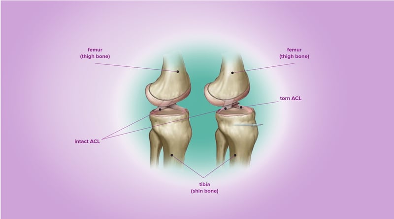
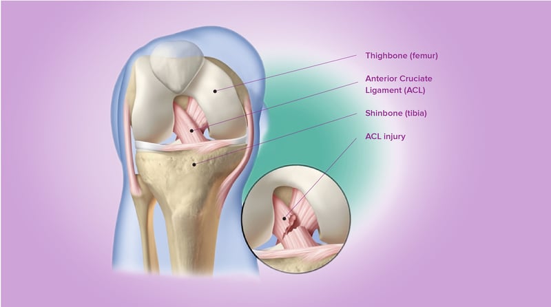
The knee joint is supported by several ligaments. These ligaments “tie” the femur (thigh bone) to the tibia (shank bone) limiting the amount of separation between the two bones at the joint. Ligaments are tough fibrous bands that are able to resist forces in tension, but not compression. Two ligaments pass through the center of the knee, the anterior cruciate ligament (ACL) and the posterior cruciate ligament (PCL). The ACL is attached to the back side of the femur and attaches to the front side of the tibia. Its purpose is to limit tibial translation in the forward direction. This can be seen more easily by viewing the side image of the knee. If the ACL is ruptured, the lower leg will be able to translate forward with respect to the rest of the knee.
Injuries to the ACL are common. They often occur during sports activities that place large forces on the knee such as suddenly stopping, jumping, or changing direction. These sports include soccer, basketball, football, tennis, downhill skiing, volleyball and gymnastics. ACL injuries are especially common in young girls playing soccer.
In cases where surgery is required, it is performed by removing the damaged ligament and replacing it with a tissue graft. The graft is obtained by removing a portion of tendon in another part of the patient’s knee or from a deceased donor. This graft will support forces on the knee joint and serve as scaffolding on which new ligament tissue can grow. Physical therapy is required following surgery to aid in the healing process to strengthen the knee and ensure the range of motion is maintained or enhanced.