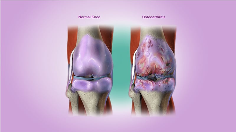
Arthrosis is another name for osteoarthritis (OA). It is the most common type of arthritis, and it caused by normal wear on joints and cartilage. Arthritis in general describes several conditions that cause inflammation in the joints. In addition to OA, two other common types of arthritis are rheumatoid arthritis and gout.

OA is classified by the severity. Lower levels are used to describe less severe conditions. For the knee joint, the Kellgren and Lawrence system is a common method of classifying the severity of OA. This is a well-established classification system proposed by Kellgren et al. in 1957 and accepted by the World Health Organization in 1961. The classification system includes five grades from zero to four.
- Grade 0: No radiographic features of OA are present. This means that no visible signs of OA are present in X-ray images of the knee.
- Grade 1: Doubtful narrowing of joint space and possible osteophytic lipping. This means that there does not appear to be narrowing of the joint space in X-ray images of the knee. The knee joint consists of cartilage-covered bone on the end of the femur, a thin pad of tough tissue, called the meniscus, and the cartilage-covered bone on the end of the tibia. Only the bone is visible on the X-ray, so a reduction in joint space indicates loss of cartilage and/or meniscus. The second condition, possible osteophytic lipping, indicates that there may be a bone growth protruding from the side of the joint. This growth is known as an osteophyte or more commonly known as a bone spur. Because osteophytes are usually caused by local inflammation, their presence is a good indicator of the existence of OA.
- Grade 2: Definite osteophytes and definite narrowing of joint space. The presence of osteophytes in X-ray images of the knee is a clear indicator of OA within the joint (or some other cause of inflammation). X-ray images should be taken while the knee is bearing weight, because body weight will force the joint closed and may show deterioration that is not present in the joint when non–weight-bearing images are taken.
- Grade 3: Moderate multiple osteophytes, definite narrowing of joint space, some sclerosis, possible deformity of bone contour. At Grade 3, there are multiple osteophytes of moderate size indicating joint inflammation. In addition, joint space narrowing is clearly visible, indicating a loss of cartilage and/or meniscus in the knee joint. Furthermore, subchondral sclerosis is visible. Subchondral sclerosis is an increase in bone thickness below the cartilage layer in response to OA. It is visible as an abnormally white region of bone along the joint line. Together with narrowing of the joint space, it is widely considered as a hallmark of OA. “Possible deformity of bone contour” indicates a potential change in overall shape of the bone structure due to the presence of OA.
- Grade 4: Large osteophytes, marked narrowing of joint space, severe sclerosis, and definite deformity of bone contour. Grade 4 is the most severe case of OA. Large osteophytes are present, indicating long-term inflammation of the joint. The joint space is clearly narrowed due to a lack of cartilage and/or meniscus protecting the joint. Severe sclerosis and definite bony deformity exists indicating the body’s response to joint deterioration.
In addition to the knee, the hip, spine, shoulder, and other joints may be classified by the Kellgren and Lawrence system. There are also many other classification systems for arthrosis. The classification systems used often depends on the joint being evaluated.
![]()